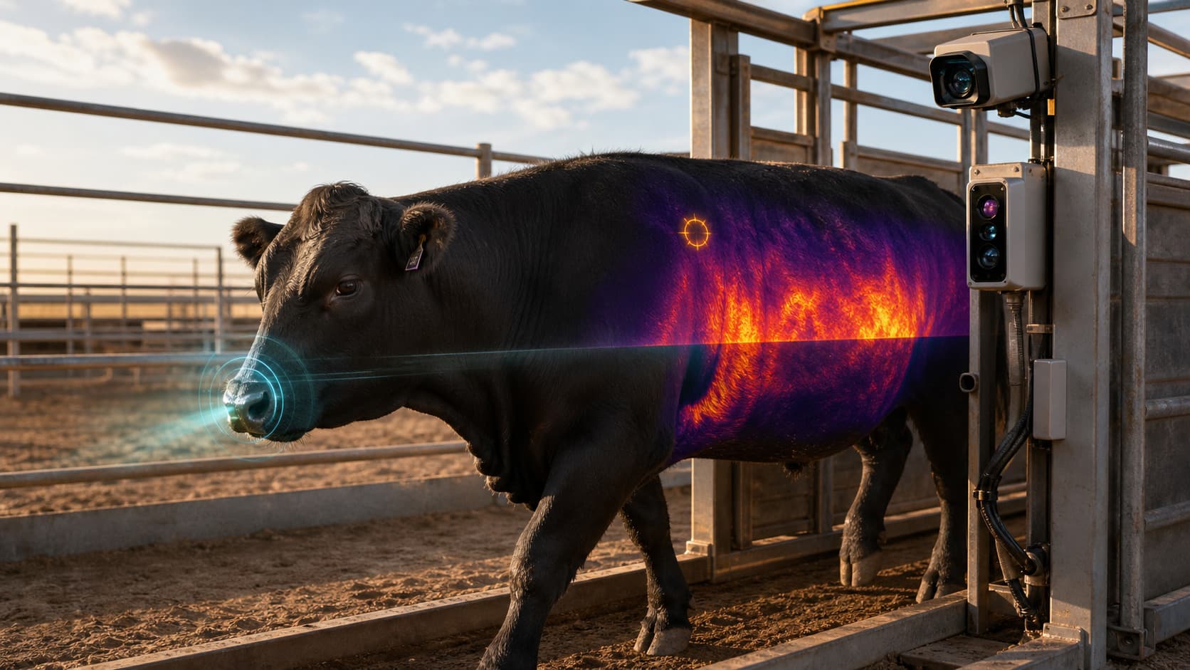
The study begins with a practical evidence question: can a controlled cattle-handling point produce consistent digital and thermal views that support repeatable wound review? The answer depends on capture geometry, synchronization, quality states, reviewer protocol, and qualified ground truth—not on a single model score.
The proposed configuration extends the Livestock Technologies chute workflow with fixed side and oblique cameras. Each passage receives a capture identifier and preserves the digital frames, thermal frames, body-region coverage, quality results, annotations, and review history.
The documented chute-capture workflow
A useful study record must be reconstructable. Research teams should be able to identify which cameras were active, which body regions were visible, how the channels were aligned, which protocol version governed review, and how an annotation changed over time.
- 01
Register the chute passage
Connect the loading record, operational identifier, timestamp, and camera station to one capture event.
- 02
Capture synchronized digital and thermal views
Record time-aligned side and oblique body views with fixed camera positions, lighting, distance, and thermal reference conditions.
- 03
Apply capture-quality gates
Record visibility, blur, obstruction, exposure, thermal range, and the body regions available for review.
- 04
Assemble versioned review evidence
Keep original frames, aligned RGB and thermal crops, annotations, reviewer decisions, protocol version, and change history together.
- 05
Measure the research protocol
Compare review labels with qualified ground truth and report coverage, disagreement, missing views, and results by site and condition.
What synchronized RGB and thermal capture contributes
Digital RGB
Document visible surface detail
Controlled color views preserve wound shape, drainage, blood, exposed tissue, coat disruption, and nearby reference tissue for review.
Thermal
Document surface-temperature patterns
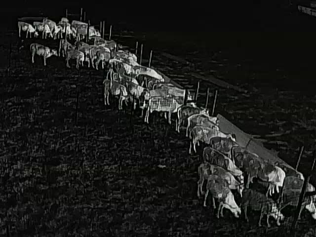
Fixed-distance thermal views preserve localized temperature asymmetry and the surrounding reference regions under recorded conditions.
Synchronized evidence
Review both channels at one body location
Time alignment and camera calibration let reviewers compare digital and thermal evidence from the same chute passage.
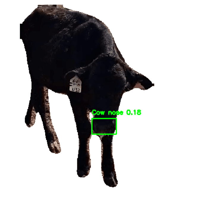
The Tuskegee University research deployment documents Livestock Technologies capture in real cattle environments using 4K digital and thermal channels. A chute study applies those modalities at closer range with controlled camera positions and a registered passage.
Versioned review evidence
The review packet should retain original files alongside calibrated derivatives. Every crop, annotation, body-location label, quality state, reviewer decision, and adjudication receives provenance. Protocol and model versions belong in the same record so later analyses can reproduce what a reviewer saw.
This structure supports blinded review, inter-reviewer agreement, correction history, and analysis of missing coverage. It also keeps the evidence useful when the research question or labeling guide evolves.
The prospective research protocol
A defensible study defines the capture and review rules before evaluating results. The protocol should specify:
- Site, cohort, handling point, camera identities, firmware, and calibration record
- Camera position, distance, angle, lighting, thermal range, and environmental context
- Body-region coverage plus explicit missing, blurred, and obstructed-view states
- Original files, aligned derivatives, annotations, reviewer identity, and protocol version
- Ground-truth method, blinded review rules, disagreement handling, and adjudication
- Prospective evaluation plan with results separated by site, coat, condition, and body region
Thermal review is strongest when distance, angle, reference regions, and ambient conditions are recorded. A peer-reviewed bovine postoperative-wound study demonstrates the value of infrared wound-temperature assessment and the need to account for measurement conditions.
Research context and source documentation
Federal and university work establishes the broader research context for multimodal wound surveillance, field confirmation, and response. The source set below anchors the study design and should be checked for current operational guidance.
Research collaboration
Design a controlled chute-capture study
Map camera geometry, synchronized capture, versioned evidence, ground truth, and prospective review with the people responsible for the research.
Primary sources
- Screwworm.gov: confirmed U.S. animal and wild-fly detections
- USDA APHIS: warning signs and suspected-case reporting
- USGS: rapid genetic testing and the role of species confirmation
- BMC Veterinary Research: infrared assessment of postoperative wound temperature in cattle
- University of Arizona Cooperative Extension: livestock inspection guide
- USDA APHIS: investment in New World screwworm preparedness projects
- USDA APHIS: Grand Challenge recommended spending plan